Tibialis Posterior Dysfunction
(Acquired Flatfoot)
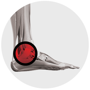
Tibialis Posterior Dysfunction (Acquired Flatfoot)
The tibialis posterior tendon is one of the major stabilising structures in the foot. It runs from the inside aspect of the ankle, from the medial malleolus, and inserts into the navicular. It supports the arch and helps to keep the foot turned inwards when walking. It can be damaged by wear and tear, and acute trauma. Initially pain is felt along the length of the tendon behind the medial malleolus but as the problem worsens, deformity can become apparent, the foot will flatten and start to turn outwards. Pain may occur on the outside part of the ankle and over time the joints can become affected and become arthritic.
Non-Operative Management
In the early stages of tibialis posterior tendon dysfunction, the use of oral analgesics, orthotics and physiotherapy are useful, and a splint may be required. The use of injections with platelet rich plasma or with cortisone are also options and Dr Rao will explain all of these to you at your consultation.
Operative Management
The surgical treatment depends upon the location and severity of the damage. In most cases the tendon itself needs to be debrided and another tendon needs to be harvested and transferred to help it work. This tendon is the FDL (flexor digitorum longus). It lies alongside the tibialis posterior at the ankle and bends the small joints of the toes. Other tendons carry out this function so the tendon is not really missed when it is used. To improve the biomechanics of the tendon transfer, the heel bone is also shifted inwards in a procedure known as a medial sliding calcaneal osteotomy.
In advanced cases the foot becomes arthritic and the patient requires a procedure known as a triple arthrodesis. Triple arthrodesis involves 3 joints, the sub-talar joint, talo-navicular joint and calcaneo-cuboid joint. Recovery from surgery does take a long time, you will be non-weight bearing for an extended period of time prior to having a boot to help you ambulate. You will also need intensive physiotherapy for a period of up to 16-20 weeks.
Risks and Complications
The general risks and complications will be discussed with you by Dr Rao. The main risks are of infection, clots and anaesthetic problems. The outcome of the surgery is good with 90%-95% of patients happy at 6 months post surgery.
Recovery Times
In terms of hospital stay:
- 3-4 nights in hospital;
- Rest, elevation for 2 weeks;
- Boot for 8 weeks;
- Time off work:
- for seated jobs, 6 weeks
- for standing jobs 14 weeks
- Foot swelling for 12-16 weeks
Meet Dr Rao
Dr Rao is a Newcastle Orthopaedic surgeon who specialises in all aspects of foot and ankle surgery. He graduated from medicine at the University of Sydney and trained as an orthopaedic surgeon in Newcastle and also in Queensland.
He also undertook further training by way of fellowship with world famous Dr Terry Saxby in Brisbane in 2008. He has also conducted further training by attending numerous courses overseas, in Thailand, the USA and right across Australia.